An MRI scan is a painless procedure that can quickly and effectively diagnose an acoustic neuroma, putting your mind at rest.
What is Acoustic Neuroma?
An acoustic neuroma (sometimes referred to as a vestibular schwannoma or acoustic schwannoma) is a benign acoustic tumour found in the ear that may negatively affect balance and hearing.
Acoustic Neuroma Symptoms
If you have an acoustic neuroma, symptoms may not be immediately apparent and can appear gradually, including such things as:
-
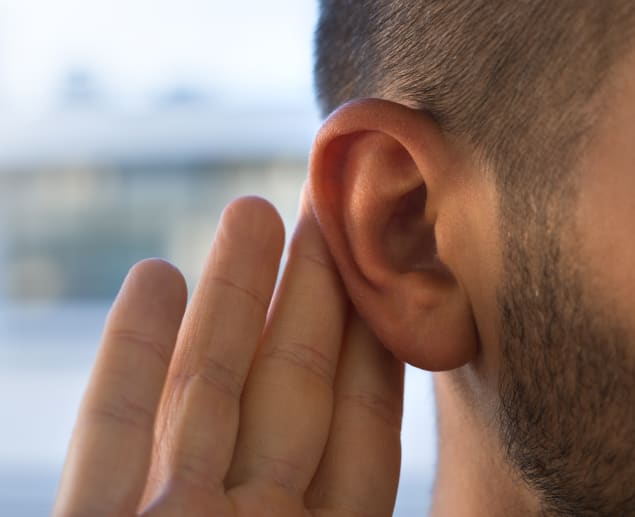
Loss of hearing
-
Tinnitus
-
Vertigo/balance problems
-
Persistent headaches
-
Blurred/double vision
-
Weakness/pain on one side of the face/facial numbness (due to compression of the facial nerve)
-
Problems with swallowing or hoarseness
Diagnosing Acoustic Neuroma
If you suspect you may have an acoustic neuroma, you may be worried. Rest assured, there are various ways of diagnosing the condition, including a hearing test (audiometry) carried out by an audiologist using tones and words.
Imaging also helps to diagnose acoustic neuroma. A magnetic resonance imaging machine (MRI) with contrast dye can detect the smallest of tumours. Computerised tomography (a CT scan) may also be used, but it can miss small tumours, which is why an MRI scan is the preferred option.
Medical History and Symptom Review
Your doctor will take into account your medical history while investigating your symptoms, some of which may be due to the cranial nerve VIII (the vestibulocochlear nerve), the brainstem or the cerebellum being compressed. This causes an increase in intracranial pressure (ICP).
Most commonly, if you have an acoustic neuroma, hearing loss will result due to reduced blood supply to the cochlear nerve. A chromosome 22 disorder can also cause problems with hearing loss. Your doctor will also examine the porus acusticus (opening to the internal acoustic canal). The facial nerve, vestibulocochlear nerve and labyrinthine artery all pass through this canal. The trigeminal nerve, which deals with sensory information of the face, can also be affected.
Depending upon the size of the non-cancerous tumour, you may notice symptoms of tinnitus, difficulty in understanding words, vertigo (dizziness) and headaches or numbness of the face. As the acoustic neuroma enlarges, the brainstem may be affected, changing the way that you walk. Knowing what to expect will make the examination less worrying for you. It may include any of the following:
Hearing Test
Your doctor will arrange a hearing test as part of the physical examination. This test will usually involve the following:
-
An evaluation of your speech
-
The use of tuning forks with varied frequencies
-
A Weber test to see if your hearing loss is bilateral or unilateral
-
A Rinne test to assess the conduction of sound through the air and bone
-
A evaluation for tinnitus, which can present as ringing or buzzing in the ears
Speech Audiometry
This is a type of hearing test that investigates how well you can hear speech and sounds. The speech audiometry test detects the presence of an acoustic neuroma by looking at:
-
The pure tone average (PTA) – how loud the sound needs to be before you can detect it.
-
The speech reception threshold (SRT) – how loud speech needs to be before you can detect it.
-
Speech discrimination (SD) – how many words you can detect in the ear at a time
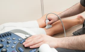
MRI Scan
An acoustic neuroma can be detected by taking a magnetic resonance imaging (MRI) scan of the brain. It makes sense to investigate symptoms early, as if treatment is required, this can be organised as soon as possible. Sometimes a contrast material, (gadolinium), will be injected during the scan to clearly show the size and location of the benign tumour. An MRI scan gives a more detailed image than a CT scan.
CT Scans
A computed tomography (CT) scan is a commonly used medical imaging procedure that uses X-rays to create a detailed picture. A computer is used to produce cross-sectional images (slices) of inside the ear, including bone, blood vessels, and soft tissue. It gives a more detailed image than an X-ray.
Brainstem Auditory Evoked Response (BAER)
A diagnostic procedure called a brainstem auditory evoked response (BAER) measures electrical activity created by the brain in response to sounds. It is used to evaluate the quality of brainstem auditory pathways, enabling detection of hearing loss and abnormalities of the nervous system.
Electronystagmography (ENG)
This is a test used to measure eye movements and the condition of the cranial nerves. Doctors use it to diagnose conditions of the inner ear, nerves, and brain, such as problems with vision, hearing, and balance, as well as dizziness and vertigo. These can occur when the vestibular nerve is affected. Your internal auditory canal (IAC) may also be examined.
Specialist Consultations
If your GP feels that you need to be seen by a specialist, you will be referred. These professionals vary by name and the area of detection for which they are responsible:
Otolaryngologist
Sometimes referred to as an ENT, the otolaryngologist treats conditions of the ears, nose, and throat and can also perform surgery on the head, neck, ears, face, nose, throat, and mouth.
Neurotologist
Responsible for the neurotology department, the neurotologist will help to diagnose, investigate, manage and rehabilitate any disorders relating to the voice as well as complex auditory issues and surgery on the skull.
Neurologist
A neurologist is a specialist who understands the layout and function of the nervous system and will diagnose, treat, and manage any disorders of the brain and nervous system (nerves, brain, and spinal cord).
Neurosurgeon
This doctor focuses on problems with the nervous system, including the nerves, spinal cord and brain. They also provide nonsurgical treatments
What Causes Acoustic Neuroma?
One question that may feature prominently is, ‘What causes acoustic neuroma?’ It is thought that the problem begins at a cellular level when a key ‘governor’ gene fails to suppress the production of Schwann cells. These cells coat nerve fibres with insulation; if not suppressed, the cells eventually grow to form a neuroma. It may also be caused by neurofibromatosis type 2 (NF2), a genetic condition that can trigger tumours to grow on nerves.
Differential Diagnosis
During your examination, doctors and specialists will attempt to rule out similar conditions with comparable symptoms. They will use a differential diagnosis process to find the cause of your symptoms and possible related conditions.
Treatment Options
A range of treatment options can be used to treat acoustic neuroma, such as medication, surgery and alternative treatments. Let’s look at some of these in more detail:
Microsurgical Removal
If the specialist decides that you need surgery to remove the acoustic neuroma, they may suggest microsurgery. This is carried out using a microscope and special instruments that allow work on delicate parts of the body.
Retrosigmoid (Suboccipital) Surgery
The surgeon will make a small incision behind the ear, opening up the skull to access the occipital bone, which is located behind the mastoid. This is referred to as retrosigmoid (suboccipital) surgery.
Translabyrinthine Surgery
Translabyrinthine surgery involves the removal of the acoustic neuroma tumour from the cerebellopontine angle, which is accessed by a small incision behind the ear.
Middle Fossa Surgery
Don’t be alarmed if your surgeon refers to you needing a middle fossa craniotomy. This is simply surgery that allows small acoustic neuromas to be treated, enabling hearing to be retained.
Radiation Therapy
Radiotherapy (radiation therapy) may be recommended by your consultant to treat the acoustic neuroma. It is a process that uses high-level radiation to kill unwanted cells. It can help reduce pain and discomfort.
Stereotactic Radiosurgery (Gamma Knife)
Gamma knife radiosurgery (stereotactic radiosurgery) is a noninvasive treatment that uses gamma radiation to treat lesions or tumours in the brain or upper spine.
Fractionated Stereotactic Radiotherapy (FSR)
Fractionated stereotactic radiotherapy (FSR) is used to treat tumours while reducing the damage to other areas. The doses of radiation are split into smaller doses and applied over several days. This ‘fractionated’ radiotherapy is a safer method that helps to reduce radiation exposure.
Cochlear Implants
Cochlear implants are used to help restore hearing function. A tiny electronic device, the implant will improve your hearing if it is being affected by the acoustic neuroma.
Supportive Treatments
If your doctor or consultant talks about providing you with supportive treatment, this refers to a type of care that will help you to feel better. This can involve such things as pain management, nutritional support from a dietician or other helpful forms of treatment.
Hearing Aids
If the medical practitioner recommends the use of a hearing aid, you don’t need to be concerned as nowadays they can be fairly unobtrusive but very powerful. If you are struggling to hear certain sounds due to having an acoustic neuroma, a hearing aid will magnify sounds so that you can hear voices, assorted sounds and music more clearly.
Vestibular Therapy
Sometimes referred to as VRT, vestibular therapy is a physical therapy that utilises a range of exercises to improve balance and eradicate dizziness that may be caused by the presence of the acoustic neuroma. The exercises are carefully designed to enable the central nervous system to function well despite any problems you may have with the inner ear.
How an MRI Scan Detects Acoustic Neuroma
An MRI scanning machine is a sophisticated piece of equipment that detects the presence of tumours, such as bilateral acoustic neuromas. It uses a magnetic field to generate three-dimensional computerised images of the soft tissue. Doctors sometimes inject the patient with a dye called gadolinium before the scan to brighten the appearance of the acoustic neuroma and make it stand out from the healthy tissue.
Why an MRI Scan is a Good Option
Having an MRI scan is a good option if your physician feels you may have an acoustic neuroma, which is a benign tumour. The images provided by the MRI scan can help diagnose whether it is present. If you suffer from claustrophobia, let the radiographer know so they can keep you comfortable. Having a scan is painless as the magnetic fields and radio waves used are undetectable by the human body.
Find an MRI Scan for Acoustic Neuroma
Finding something wrong with your body can be worrying, which is why early detection by an MRI scan is so helpful. If you are concerned that you may have a tumour, such as an acoustic neuroma, but the NHS waiting list is long, lessen your concern by contacting us to arrange a private MRI scan. Our friendly clinicians also provide consultations if you wish to find out more about whether a scan is right for you.
Get in touch with our friendly customer service team today and put your mind at rest by booking a private MRI scan.
Sources
Acoustic neuroma - Diagnosis and treatment - Mayo Clinic. (n.d.). https://www.mayoclinic.org/diseases-conditions/acoustic-neuroma/diagnosis-treatment/drc-20356132
Acoustic Neuroma (Vestibular Schwannoma). (2023, March 6). Johns Hopkins Medicine. https://www.hopkinsmedicine.org/health/conditions-and-diseases/brain-tumor/vestibular-schwannoma
BAER - brainstem auditory evoked response. (n.d.). Mount Sinai Health System. https://www.mountsinai.org/health-library/tests/baer-brainstem-auditory-evoked-response
Benefits and risks. (2017, December 9). U.S. Food And Drug Administration. https://www.fda.gov/radiation-emitting-products/mri-magnetic-resonance-imaging/benefits-and-risks
Canty, P. (1978). Speech audiometry in acoustic neuroma. The Journal of Laryngology & Otology, 92(10), 843–851. https://doi.org/10.1017/s0022215100086217
Cochlear implants. (2024, June 13). NIDCD. https://www.nidcd.nih.gov/health/cochlear-implants
Cohen-Gadol, A., MD. (2024). Overview of acoustic neuroma. www.aaroncohen-gadol.com. https://www.aaroncohen-gadol.com/en/patients/acoustic-neuromas/overview
Diagnosing acoustic neuroma. (n.d.). NYU Langone Health. https://nyulangone.org/conditions/acoustic-neuroma/diagnosis
Electronystagmography (ENG). (2021, August 8). Johns Hopkins Medicine. https://www.hopkinsmedicine.org/health/treatment-tests-and-therapies/electronystagmography-eng
Gamma Knife radiosurgery. (2024, May 6). Johns Hopkins Medicine. https://www.hopkinsmedicine.org/health/treatment-tests-and-therapies/gamma-knife-radiosurgery
Greene, J., & Al-Dhahir, M. A. (2023a, August 17). Acoustic neuroma. StatPearls - NCBI Bookshelf. https://www.ncbi.nlm.nih.gov/books/NBK470177/
Han, B. I., Song, H. S., & Kim, J. S. (2011). Vestibular Rehabilitation therapy: Review of indications, mechanisms, and key exercises. Journal of Clinical Neurology, 7(4), 184. https://doi.org/10.3988/jcn.2011.7.4.184
Hearing aids. (2022, October 11). NIDCD. https://www.nidcd.nih.gov/health/hearing-aids
NCI Dictionary of Cancer Terms. (n.d.). Cancer.gov. https://www.cancer.gov/publications/dictionaries/cancer-terms/def/supportive-care
Professional, C. C. M. (2024a, May 1). Differential diagnosis. Cleveland Clinic. https://my.clevelandclinic.org/health/diagnostics/22327-differential-diagnosis
Professional, C. C. M. (2024b, May 1). Microsurgery. Cleveland Clinic. https://my.clevelandclinic.org/health/treatments/microsurgery
Professional, C. C. M. (2024c, May 1). Neurologist. Cleveland Clinic. https://my.clevelandclinic.org/health/articles/22277-neurologist
Professional, C. C. M. (2024d, May 1). Neurosurgeon. Cleveland Clinic. https://my.clevelandclinic.org/health/articles/22364-neurosurgeon
Professional, C. C. M. (2024e, August 20). Otolaryngologist. Cleveland Clinic. https://my.clevelandclinic.org/health/articles/24635-otolaryngologist
Radiation therapy - Mayo Clinic. (n.d.). https://www.mayoclinic.org/tests-procedures/radiation-therapy/about/pac-20385162
RNID. (2023a, December 18). Acoustic neuroma - RNID. https://rnid.org.uk/information-and-support/hearing-loss/types-of-hearing-loss-and-deafness/acoustic-neuroma/
Showalter, T., Werner-Wasik, M., Curran, W., Friedman, D., Machtay, M., Evans, J., & Andrews, D. (n.d.). Fractionated Stereotactic Radiotherapy (FSR) and Stereotactic Radiosurgery (SRS) for the Treatment of Nonacoustic Cranial Nerve Schwannomas. International Journal of Radiation Oncology*Biology*Physics, 69(3), S164. https://doi.org/10.1016/j.ijrobp.2007.07.296
Surgery for acoustic neuroma. (n.d.). NYU Langone Health. https://nyulangone.org/conditions/acoustic-neuroma/treatments/surgery-for-acoustic-neuroma
The Brain Tumour Charity. (2024, May 28). Acoustic neuroma | The Brain Tumour Charity. https://www.thebraintumourcharity.org/brain-tumour-diagnosis-treatment/types-of-brain-tumour-adult/acoustic-neuroma-vestibular-schwannoma/
Translabyrinthine Approach to Acoustic Neuroma Dallas, Texas. (n.d.). http://www.centerforacousticneuroma.com/treatment-translabyrinthine-approach.html
Treatment definition. (n.d.). https://hsc.unm.edu/about/administrative-departments/privacy-office/treatment-definition
Waterval, J., Stokroos, R., & Dings, J. (2014). Middle Fossa approach. Otorhinolaryngology Clinics - an International Journal, 6(4), 19–24. https://doi.org/10.5005/aijoc-6-4-19
NHS. (2022, August 5). MRI scan. nhs.uk. https://www.nhs.uk/conditions/mri-scan/