Key takeaway:
-
Medical scans produce detailed images of various parts of the body.
-
Scan images are used by doctors to diagnose the cause of symptoms such as pain, swelling and abnormal bleeding.
-
There are different types of scans, including MRI, CT, X-ray and ultrasound.
-
Different medical scans are used to examine various parts of the body and internal structures, such as bones, organs, soft tissues, and blood vessels.
-
The right scan for you will depend on your symptoms and medical history.
Why Medical Imaging Scans Are Used
Doctors and other medical specialists use medical scans to see inside the body without having to perform surgery or invasive procedures.
Medical scans create images of the internal structures of the body. They help doctors diagnose the causes of symptoms such as pain, inflammation, swelling and abnormal bleeding. They’re also used to monitor disease progression and plan treatments.
Some types of scans require a contrast dye injected into a vein, but generally, medical scans are painless and non-invasive.
Main Types of Medical Imaging Scans
There are different types of scans used to create images of the inside of the body: MRI scans, CT scans, X-rays and ultrasound scans. Your doctor will advise you on the appropriate scan based on your symptoms, medical history and the parts of the body requiring detailed images.
MRI Scans
MRI stands for Magnetic Resonance Imaging. An MRI scan creates three-dimensional images of the body's internal structures. They generally take longer than CT scans, are noisy and take place inside an enclosed space, but unlike CT scans, they don’t use radiation.
What Are MRI Scans?
An MRI scanner uses radio waves and a strong magnetic field to create detailed images of the inside of the body. Patients are required to lie very still on a table that slides into a doughnut-shaped, tunnel-like MRI machine while detailed images are taken.
What are MRI Scans Used For?
MRI scans are used to diagnose and monitor conditions affecting the bones, joints, soft tissues (including muscles, tendons and ligaments), organs, nerves, spinal cord and blood vessels.
They’re particularly useful for evaluating soft tissue injuries, joint strains and sprains, back pain, persistent headaches and neurological symptoms. An MRI scan is also used to help diagnose certain types of cancer. MRI images show very subtle differences between healthy and abnormal tissue that might not be detected on a CT scan.
Types of MRI Scan
-
Standard MRI - a ‘closed’ MRI taken in a long, doughnut-shaped, tunnel-like machine that produces highly detailed images.
-
Contrast MRI - a standard MRI with the addition of a contrast dye, injected into a vein to help better highlight blood vessels, inflammation and potential cancerous tumours.
-
Open MRI - an MRI scan taken in a newer, smaller type of open MRI machine that’s potentially easier for claustrophobic patients.
-
Functional MRI - an MRI that measures blood flow and activity in the brain during functions such as movement, speech and recall.
-
Diffusion-Weighted Imaging - an MRI scan that measures the movement of water molecules through tissues, helpful for identifying strokes and cancerous tumours.
-
Diffusion Tensor Imaging - an advanced MRI technique that measures the movement of water molecules through the brain to assess brain connectivity, diagnose strokes and brain tumours and plan surgical procedures.
-
MRI Arthrogram - specialised MRI scanning after a contrast dye has been injected into a joint space, often in the shoulder or hip, to help diagnose soft tissue tears and damage.
-
MR Angiography - highlights the arteries to assess for narrowing, blockages and aneurysms.
-
MR Venography - highlights the veins to assess for narrowing, blockages and blood clots.
-
MR Spectroscopy - advanced MRI scanning that measures chemicals and metabolites in the brain tissue, used in the diagnosis of strokes and brain tumours.
-
Cardiac MRI - an MRI scan focused on the heart, including the valves, chambers and blood vessels, to evaluate damage and disease.
-
Dynamic Contrast-Enhanced MRI - a type of contrast MRI used to assess blood flow, usually through the heart and brain.
CT Scans
CT stands for Computed Tomography. A CT scan creates three-dimensional, cross-sectional images of the inside of the body. They’re quick and often used in emergency situations as well as in routine examinations. CT scans use a low dose of radiation.
What Are CT Scans?
CT scanners use X-ray technology to create detailed images of the internal structures of the body. Quicker than an MRI scan, patients lie on a bed that moves into a short, round CT scanner as images are taken. They can feel less claustrophobic than traditional MRI scanners.
What are CT Scans Used For?
CT scans are used to diagnose and monitor conditions affecting the bones, soft tissues, organs and blood vessels.
They’re particularly useful for assessing bone fractures, including in the skull, that might not be picked up on a standard X-ray, and internal bleeding. CT scans are also used to detect blood clots and embolisms, as well as damaged or diseased organs, including the lungs and the organs of the abdomen and pelvis.
A CT scan can also be used to diagnose some forms of cancer, sometimes in conjunction with a positron emission tomography, or PET scan.
Types of CT Scan
-
Standard CT - a routine CT scan that focuses on the whole body or specific body parts.
-
Contrast CT - a standard CT with the addition of a contrast dye injected into a vein to better highlight the blood vessels, inflammation and potential cancerous tumours.
-
Low-Dose CT - specialised, minimal radiation CT scanning, often used to scan the lungs of high-risk patients such as long-term or former smokers and the elderly, to help diagnose lung cancer.
-
High-Resolution CT - highly specific CT scanning of the lungs to help diagnose lung cancer and lung disease.
-
CT Angiogram - a CT scan that highlights the arteries to diagnose narrowing, blockages and aneurysms.
-
CT Venography - a CT scan that highlights the veins to diagnose narrowing, blockages and blood clots.
-
Cardiac CT - a CT scan that focuses on the heart to help in the diagnosis of plaque buildup, blockages and aneurysms.
-
CT Colonography - a non-invasive ‘virtual’ colonoscopy that scans the large bowel looking for polyps and tumours.
-
Dual-Energy CT - a CT scan that uses two separate X-ray beams to separate tissues on the image and create a clearer picture.
-
CT Calcium Score - a specialised CT scan of the heart and main arteries to measure calcium buildup, a sign of potential heart disease.
X-Ray Scans
X-rays create images of the inside of the body using electromagnetic radiation. They’re quick, and patients don’t need to enter a scanning machine, but they use radiation at a low dose.
What Are X-Rays?
During an X-ray, electromagnetic radiation passes through the body onto an X-ray film, which captures an image as a shadow. The denser the tissue type, such as bones and teeth, the lighter the shadow. Soft tissues appear as darker shadows on an X-ray image.
What are X-Rays Used For?
X-rays are often a “first-line” scan, used to reveal significant bone fractures. They’re also used to diagnose dislocations, joint narrowing, dental problems and pneumonia, as well as to identify foreign objects within the body.
An X-ray shows soft tissues in less detail than CT or MRI scans, but it can reveal abnormal masses in the organs or bones that may require further investigation.
Types of X-Ray
-
Digital X-Ray - a standard X-ray performed using digital sensors to create images, rather than the older style X-ray film.
-
Computed Radiography - a lower dose, digital X-ray that creates latent, or hidden, images on a reusable plate, rather than sensors or film, that are then converted into a digital image.
-
Fluoroscopy - an X-ray taken after the administration of a contrast dye into a vein, to track its movement through the body, usually specifically the heart, to check for blockages and to guide stent placement.
-
Contrast X-Ray - X-ray images taken after a patient ingests barium, either as a liquid or via enema, or after barium is injected into a vein, to highlight problems affecting the large intestines, stomach or kidneys.
-
Mammography - specialised, low-dose X-ray scanning of the breasts to detect cancer or as a routine screening test for breast cancer.
-
Tomosynthesis - a specialised type of breast screening that produces highly detailed images, especially useful in patients with dense breast tissue.
-
Mobile X-Ray - X-rays taken using a smaller, portable X-ray machine, useful if a patient is in critical care, immobile or is too unwell to move to an X-ray department.
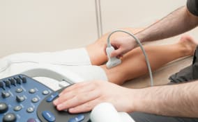
Ultrasound Scans
Ultrasound scans, also called sonography, create images of parts of the body in real time using sound waves and don’t use radiation.
What are Ultrasound Scans?
During an ultrasound, patients lie on a medical bed while an ultrasound probe called a transducer is moved over the skin. They create moving images used to assess internal organs in real time.
What are Ultrasound Scans Used For?
Ultrasound scans are used to assess organs and soft tissues. They’re routinely used to diagnose conditions such as gallstones and kidney stones, to help diagnose conditions affecting the eyes, thyroid gland, liver, bladder, ovaries and uterus, and to check blood flow through the organs. Ultrasound is also used to monitor health and development during pregnancy.
Types of Ultrasound Scan
-
Standard Ultrasound - a routine ultrasound scan using a probe called a transducer applied to the skin externally, transvaginally (inside the vagina) or transrectally (inside the rectum).
-
Doppler Ultrasound - a specialised type of ultrasound that measures blood flow through the blood vessels.
-
Colour Doppler Ultrasound - a Doppler ultrasound that uses colour to represent and highlight the speed and direction of blood flow.
-
Power Doppler Ultrasound - a highly sensitive Doppler ultrasound that can detect very slow blood flow or blood flow through very small blood vessels and capillaries.
-
Spectral Doppler Ultrasound - a specialised Doppler ultrasound that produces a waveform, or graphical representation of blood flow and speed over time.
-
3D Ultrasound - an advanced pregnancy ultrasound that creates highly detailed, three-dimensional images of an unborn baby.
-
4D Ultrasound - a further advanced pregnancy ultrasound that creates a moving video of three-dimensional images of an unborn baby.
-
Contrast-Enhanced Ultrasound - a specialised ultrasound taken after a contrast agent containing microbubbles has been injected into a vein to better highlight blood flow, specifically through the organs, including the pancreas, liver, kidneys and spleen.
-
Elastography - a type of ultrasound that measures the elasticity or ‘stiffness’ of certain tissue, including liver, thyroid, prostate and breast tissue.
-
Fibroscan - a specialised type of elastography ultrasound that measures the stiffness of the liver, which might indicate scarring and liver diseases.
Other Scans
-
Gynaecology Scan - an internal ultrasound called a transvaginal ultrasound that's used to diagnose the cause of pelvic pain and abnormal bleeding, and to assess aspects of fertility.
- Health Scans - screening options and proactive health checks
-
Heart Health Scan - a type of ultrasound scan called an echocardiogram that assesses the health of the heart and the surrounding blood vessels.
-
MRI Stress Test - an MRI of the heart performed after gentle treadmill exercise or after a drug that makes the heart work harder, to analyse how the heart works under stress.
-
Urology Scan - an ultrasound of the prostate, kidney or bladder to help diagnose an enlarged prostate, prostate cancer, urinary tract issues and cancer.
Which Medical Imaging Scan Do I Need?
Your doctor will recommend the types of scan that are best for you, based on your symptoms, where in your body you’re experiencing them and your medical history.
Are Medical Imaging Scans Safe?
Yes, all types of scans are considered safe for most people. However, if you have a metal implant, prosthesis, mesh, stent or medical clip inside your body, you may not be suitable for an MRI scan as magnetic metal is dangerous inside an MRI scanner. Some scans may be unsuitable in the first trimester of pregnancy. Your doctor will advise you.
How To Book A Medical Imaging Scan
Booking a medical scan through Scan.com is easy. Simply choose a location and a time that suits you. We’ll call you to ask a few questions about your symptoms, and then we’ll organise a referral letter for your chosen clinic. You can then attend your scan at your chosen time.
Wth over 250 locations to choose from, we’re the UK’s largest imaging provider, and we’re rated ‘Excellent’ on Trustpilot. Jump the queues and NHS waiting lists, and get your results in days.
If you’re unsure what scan you need, book a consultation with one of our clinicians for expert scan advice and a no-obligation scan referral if an imaging test is recommended.