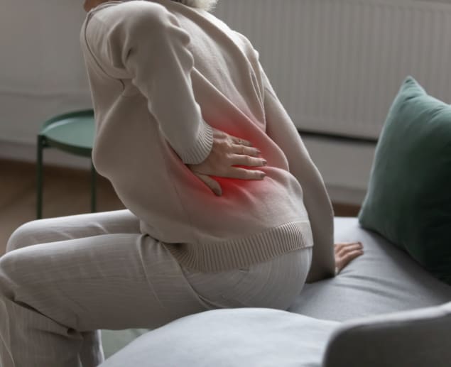

If you're experiencing persistent back pain or symptoms that suggest spinal stenosis, you might be exploring options for diagnosis and treatment.
This comprehensive article explores spinal stenosis in detail, including what it is, its symptoms, causes, stages, types, and available treatment options. Most importantly, we discuss how a spinal stenosis MRI scan, the preferred diagnostic imaging option, offers a clear and detailed view of your spine to allow investigation and an accurate diagnosis.
What is spinal canal stenosis?
Spinal canal stenosis is a medical condition where the space within your spinal canal becomes smaller, potentially placing pressure on the spinal cord and nerve roots (the first part of nerves where they branch out of the spinal canal).
Your spinal cord is a bundle of nerve fibres that run from the base of your skull to just above your hips. Just as your brain is protected by the skull, your spinal cord is safeguarded by up to 24 stacked bones called vertebrae or backbones.
These vertebrae have a hole at their centre (called the vertebral foramen), which forms a hollow tube around the spinal cord. On either side of the spine, where two adjacent vertebrae join, are openings (called the neural foramina), which allow nerves to exit and branch out to various parts of your body.
Additionally, between each vertebra are spongy tissues (called intervertebral discs) that provide cushioning, maintain stability, and allow frictionless movement.
The space or room where your spinal cord is enclosed is called the spinal canal.
The word stenosis means narrowing or constriction of any bodily passage.
Spinal canal stenosis is prevalent among older adults aged 50 years and up, resulting from thickening or swelling of facet joints (the special joints that allow the vertebrae to move against each other) in response to long-term stress or an ageing-related condition known as osteoarthritis. It can also be seen in young people and may be caused by any of the following:
-
Bone spurs (bony overgrowth).
-
Spinal cysts or tumours.
-
Fracture or injury of spinal bone(s).
-
Disc problems like herniated or slipped discs caused by degeneration or trauma.
-
Abnormal spine alignment (e.g., scoliosis and kyphosis).
-
Congenital (present at birth) bone disorders.
-
Certain bone diseases like Paget disease and rheumatoid arthritis.
Also, if you are born with a narrow spinal canal, you may be at risk of developing spinal stenosis.
What are some symptoms of spinal stenosis?
Depending on the part of the spine affected and severity of the stenosis, you may experience the following spinal stenosis symptoms:
-
Pain in the back or neck.
-
Sciatica (a nerve injury characterised by burning pain that radiates from the buttocks down to the legs or feet).
-
Pain that worsens with downhill walking but lessens with uphill walking.
-
Pain that lessens when you bend forward or sit.
-
Numbness or tingling in the arms, hands, legs, or feet.
-
Declining fine motor skills, particularly the use of the hands for buttoning a shirt, writing, and typing.
-
Muscle weakness, heaviness, or cramping in the lower limbs.
-
Abnormal reflexes.
-
Poor balance and coordination.
In its early stages, spinal stenosis often causes no symptoms, but as the spinal canal continues to narrow and compress or pinch nerves, symptoms can develop and worsen over time. In severe cases, it may cause problems with bladder and bowel control.
How does spinal stenosis affect the bowels and bladder?
The cauda equina is a group of nerves and nerve roots that emerge from the end of your spinal cord, where your lower back bones are located. They provide motor (movements) and sensory (ability to feel sensations) functions to your legs, bladder, anus, and perineum.
When spinal stenosis occurs in the lower back and compresses or pinches the cauda equina so much that it becomes irritated, its functions may be disrupted, thereby resulting in a condition known as cauda equina syndrome (CES). Bladder and bowel dysfunction is a common symptom of this serious condition.
With CES, you may experience a loss of the sensation or urgency to use the bathroom when your bladder or bowel is full or loss of bladder and anal muscle control, which may cause incontinence.
Other symptoms may include:
- Severe lower back pain
- Difficulty walking
- Sciatica
- Decreased sensation in the lower extremities
- Sexual dysfunction in men
What are the different types of spinal stenosis, and where can they occur?
There are two main types of spinal stenosis, namely:
-
Foraminal stenosis: The openings where nerve roots leave the spinal cord are called neural foramina, so foraminal stenosis refers to the narrowing of one or more of these openings, resulting in the compression of spinal nerves that pass through. It is the most commonly occurring type of stenosis and is also referred to as lateral stenosis since the narrowing occurs laterally (at the sides) of the spinal canal.
-
Central canal stenosis: Earlier, we also mentioned that the hollow space formed by the vertebral bones to enclose and protect the spinal cord is called the spinal canal. Central canal stenosis occurs when this space becomes smaller, placing direct pressure on the spinal cord.
Both foraminal and central canal stenosis can coexist and may manifest anywhere along the spine. However, they most commonly affect two regions, namely:
-
Neck (cervical stenosis): Your neck has seven stacked bones (vertebrae) called the cervical spine. Labelled C1 to C7, the cervical vertebrae are the smallest and lightest bones in the spine.
-
Lower back (lumbar stenosis): The bones in your lower back—five of them labelled L1 to L5—are known as the lumbar spine. They are the largest and thickest bones of the spine.
Though rare, stenosis can also occur in the upper/middle back (thoracic stenosis), affecting any of the 12 bones known as T1 to T12 of the thoracic spine.
What are the 4 stages of spinal stenosis?
The four stages of spinal stenosis are not based on the severity of stenosis symptoms but on imaging scan results. Healthcare providers use these stages—or, as otherwise known, grades—to describe the degree of narrowing in the spinal canal or foraminal openings and the amount of pressure exerted on the spinal cord or nerve roots as a result. They include:
-
Stage 0 or no spinal stenosis - the spinal cord and nerves pass, is normal in size, and there’s no compression of neural structures.
-
Stage 1 or mild stenosis - Here, degeneration has likely just kicked off, and the spinal canal or nerve openings are starting to narrow, but without any compression. At this stage, it is hard to tell that stenosis is occurring without an actual detailed view of the spine, as there aren’t usually presenting symptoms.
-
Stage 2 or moderate stenosis - The stenosis has progressed, causing the spinal canal and/or nerve openings to become smaller and more restrictive. On scan images, the spinal cord and nerve roots may still maintain a normal appearance. However, if symptoms of stenosis are present, it indicates significant nerve impingement, meaning a nerve or nerve root is being compressed or pinched.
-
Stage 3 or severe stenosis - At this stage, the spinal canal or nerve openings become severely restrictive, allowing nearby bones, discs, or ligaments to press on the spinal cord, nerves, or nerve roots. This heightened pressure may lead to structural changes or deformities, which can only be seen on scan images.
For instance, in severe lumbar central canal stenosis, the cauda equina may appear clumped, as though they are a single bundle, thickened, and displaced. Similarly, in severe lumbar foraminal stenosis, one or more cauda equina nerve roots may collapse, displaying changes such as oedema (swelling), atrophy (shrinkage), flattening, or thickening.
What are the final stages of spinal stenosis?
The symptoms of spinal stenosis worsen greatly in the final stages of the condition, to the extent that if conservative treatments fail to improve a person’s symptoms, they are considered to have severe (stage 3) stenosis.
A person may experience difficulty standing or walking short distances, and symptoms such as lower back or neck pain, numbness and tingling in the extremities, muscle cramping or weakness, and more, all of which can make it challenging to perform day-to-day activities.
Saddle anaesthesia (loss of sensation in the areas that would come into contact with a saddle when riding a horse) accompanied by progressive leg weakness and loss of function in the bowel and bladder could indicate a serious condition called cauda equina syndrome, a medical emergency.
Without prompt and effective treatment, permanent nerve damage can occur, leading to major physical and emotional consequences, such as permanent paralysis and depression.
How is spinal stenosis diagnosed?
The diagnosis of spinal stenosis usually begins with a complete medical history review. Your healthcare provider will ask questions about your unique symptoms, including when you first noticed them, how they feel, and whether they are aggravated or alleviated by certain actions, as well as if you have an existing medical condition or past injuries.
Next, you will undergo a physical exam where your provider may move their hands along the different regions of your spine and ask you to make some movements to assess your pain level, ability to feel sensations, reflexes, and muscle strength.
Depending on the severity of your symptoms, if your provider suspects serious nerve injury, they might proceed with a nerve function test like electromyography (EMG) or nerve conduction studies (NCS) to check the speed, strength, and consistency of nerve impulses in specific muscles when stimulated and at rest.
Finally, an imaging test will be ordered to get a good look at what's happening inside your spinal canal and rule out or confirm suspected diagnoses. The gold standard test for diagnosing spinal stenosis is a magnetic resonance imaging (MRI) scan.
A spinal stenosis MRI is a non-invasive, painless procedure that uses a powerful magnetic field and high-frequency radio waves to create highly detailed, cross-sectional images of your spine. Not only is an MRI able to visualise the spinal bones, but it can also show the intervertebral discs, muscles, fat, ligaments, spinal cord, nerve roots, and blood vessels, which allows it to identify the location, extent or stage, and cause(s) of the stenosis.
Alternative diagnostic imaging tests to the MRI for spinal stenosis include:
-
X-rays: An X-ray scan uses low-dose radiation to visualise bony abnormalities causing your spinal canal to narrow.
-
Computed tomography (CT) scan: A CT scan uses a computer to transform x-ray images taken from different angles into more detailed, cross-sectional images of your spine. It is typically used in situations where an MRI is contraindicated (i.e., not suitable for you).
A CT myelogram uses a contrast dye to enhance the visibility of your spinal cord, nerves, and other soft tissues that would easily be seen on an MRI without contrast.
What does spinal stenosis look like on MRI?
A spinal stenosis MRI shows two different pictures of the spine:
-
Cross-sectional or axial view, which looks like the spine is sliced in half from side to side
-
Sagittal view, which looks like the spine is cut in half from top to bottom.
In this view, the grey-coloured spinal cord is surrounded by a bright or white space, which is the thecal sac and a cushion of fluid protecting it (cerebrospinal fluid).
On the left side, the stacked spinal bones will be visible with darker grey discs between them and the right side, bony projections of each vertebra are visible. The white-coloured neural foramina (nerve root openings) can also have tiny, grey, circular dots in the middle, showing where the nerve roots are.
If spinal stenosis is present, the white space around the grey tube will appear smaller. This happens because an abnormality—which will also be seen in the image—is pushing into the space from either side, making it tighter and smaller than it should be.
In severe spinal stenosis MRI images, the spinal cord may appear pressed on both sides. On one side, there could be a worn-out disc or a fractured bone. On the other, there might be a thickened ligament, bone spur or, rarely, a bone tumour. At the exact location of the stenosis, you might not see any cerebrospinal fluid at all—it's all squeezed out.
Degenerative spinal stenosis MRI images commonly highlight issues with intervertebral discs, such as reduced height and disc bulging, slippage or herniation. Sometimes, it's not just the discs causing trouble – it may be bony overgrowths (bone spurs) or other parts of the spine that thicken or enlarge.
How is spinal stenosis treated?
How do you fix spinal stenosis without surgery?
Effective non-surgical treatments for spinal stenosis can include:
-
Light exercise and physical therapy - A physical therapist will create a personalised program consisting of a range of stretching, range-of-motion, and strengthening exercises tailored to the diagnosis made by your healthcare provider. These exercises will help strengthen and tone your back, core (abdominal), and leg muscles, stabilise your spine, and improve your flexibility, posture, and balance. You'll experience significant relief from stenosis symptoms and enjoy more fluid movements, enabling you to engage more comfortably in daily activities. Your physical therapist might also recommend light exercises to do daily outside of therapy sessions. These workouts can help reduce strain on your spine, especially if you are overweight.
-
Medication - This may involve the use of over-the-counter (OTC) painkillers, prescribed non-steroidal anti-inflammatory drugs (NSAIDs) or epidural steroids (also known as guided corticosteroid injections) to help reduce irritation, inflammation, swelling, and pain. Anti-seizure drugs such as gabapentin might also be prescribed if there is pain from a damaged nerve(s).
-
Remedies - such as heat or cold therapy, massage therapy, acupuncture, chiropractic care, and yoga may also add to ease painful symptoms in people with mild to moderate spinal stenosis.
Surgery for spinal stenosis
In severe cases of spinal stenosis, surgery might be necessary to decompress the spine. There are different types of surgery for spinal stenosis, including:
-
Laminectomy - Involves the removal of the lamina (back portion) of the affected spinal bone and the exact cause (e.g., thickened ligament, bone spur, or herniated disc) of the stenosis.
-
Laminotomy or partial laminectomy - Involves the removal of a small part of the lamina to create room in the spinal canal and relieve any pressure in the specific spot.
-
Laminoplasty - A laminotomy procedure performed only on the neck (cervical stenosis).
-
Foraminotomy - Involves the removal of tissues or bones that are compressing a nerve root as it exits the spinal canal through its designated opening (neural foramen).
What is the newest treatment for spinal stenosis?
One of the newest treatments for spinal stenosis is stem cell therapy. This regenerative medicine is not new in the space, but it has been gaining more traction for its increasing effectiveness.
Stem cells are a special type of cell capable of developing into many different types of important cells in the body. Stem cell therapy for spinal stenosis may involve the extraction of mesenchymal stem cells from the bone marrow and injecting them into the affected area to promote tissue regeneration and reduce inflammation, potentially alleviating symptoms and improving mobility.
Another fresh and innovative treatment for spinal stenosis is platelet-rich plasma (PRP) therapy. Unlike stem cell therapy, PRP involves isolating immune cells called platelets from a person's blood and injecting them into the spinal injury site. These platelets contain growth factors that stimulate tissue repair and accelerate healing, offering another promising approach in regenerative medicine for spinal stenosis.
Conclusion
An MRI spinal stenosis scan should be your go-to diagnostic imaging test if you are experiencing back pain or other symptoms of stenosis. It can accurately identify the presence of stenosis, the specific location of nerve compression, its severity, and underlying causes. This information will guide your healthcare provider in creating an effective, personalised treatment plan.
Ready to take the next step? Book a private spinal stenosis MRI scan near you today! Scan.com is the UK’s largest imaging network, with over 200+ scanning centres nationwide. Booking with us is as easy as browsing through locations near you, comparing prices, checking for the earliest appointment time, and hitting the ‘book now’ button. No GP referral needed. No NHS waiting lists.
Your results, which include an interactive, easy-to-understand radiologist report and spinal stenosis MRI images, will be prepared quickly after your scan. Also, an expert clinician will be available to explain any adverse findings and advise you on the next steps.
If you are unsure whether an MRI is the right step for you, consider booking a consultation for £50 to speak with an expert clinician. They will review your medical history, provide tailored information about the benefits and risks of the available options, and offer a no-obligation referral to the most suitable scan.
Sources:
-
Bjerke, M., & Bjerke, B. (n.d.). Types of Spinal Stenosis. Spine-health. Retrieved May 24, 2024, from https://www.spine-health.com/conditions/spinal-stenosis/types-spinal-stenosis
-
Deng, F. (2023, November 12). Lumbar foraminal stenosis | Radiology Reference Article. Radiopaedia. Retrieved May 27, 2024, from https://radiopaedia.org/articles/lumbar-foraminal-stenosis
-
Dugdale, D. C., & Conaway, B. (n.d.). Spinal Stenosis – Symptoms and Causes. Penn Medicine. Retrieved May 24, 2024, from https://www.pennmedicine.org/for-patients-and-visitors/patient-information/conditions-treated-a-to-z/spinal-stenosis
-
Electromyography (EMG) and Nerve Conduction Studies. (n.d.). MedlinePlus. Retrieved May 24, 2024, from https://medlineplus.gov/lab-tests/electromyography-emg-and-nerve-conduction-studies/
-
Lee, J. W., & Seo, J. (2023, March). Magnetic Resonance Imaging Grading Systems for Central Canal and Neural Foraminal Stenosis of the Lumbar and Cervical Spines With a Focus on the Lee Grading System. NCBI. Retrieved May 24, 2024, from https://www.ncbi.nlm.nih.gov/pmc/articles/PMC9971835/
-
Lumbar Spinal Stenosis. (n.d.). Johns Hopkins Medicine. Retrieved May 24, 2024, from https://www.hopkinsmedicine.org/health/conditions-and-diseases/lumbar-spinal-stenosis
-
MRI grading of spinal stenosis is not associated with the severity of low back pain in patients with lumbar spinal stenosis. (n.d.). NCBI. Retrieved May 24, 2024, from https://www.ncbi.nlm.nih.gov/pmc/articles/PMC9465904/
-
A Practical MRI Grading System for Lumbar Foraminal Stenosis. (2010, April). American Journal of Roentgenology. Retrieved May 24, 2024, from https://ajronline.org/doi/full/10.2214/AJR.09.2772
-
Rider, L. S., & Marra, E. M. (n.d.). Cauda Equina and Conus Medullaris Syndromes - StatPearls. NCBI. Retrieved May 24, 2024, from https://www.ncbi.nlm.nih.gov/books/NBK537200/
-
Spinal stenosis. (n.d.). Wikipedia. Retrieved May 24, 2024, from https://en.wikipedia.org/wiki/Spinal_stenosis
-
Spinal Stenosis Diagnosis & Treatment - NYC | Columbia Neurosurgery in New York City. (n.d.). Columbia Neurosurgery in New York City. Retrieved May 24, 2024, from https://www.neurosurgery.columbia.edu/patient-care/conditions/spinal-stenosis
-
Spinal Stenosis Symptoms & Causes | NIAMS. (n.d.). National Institute of Arthritis and Musculoskeletal and Skin Diseases. Retrieved May 24, 2024, from https://www.niams.nih.gov/health-topics/spinal-stenosis
Sritharan, K., Chamoli, U., Kuan, J., & Diwan, A. D. (2019, September 26). Assessment of degenerative cervical stenosis on T2-weighted MR imaging: sensitivity to change and reliability of mid-sagittal and axial plane metrics. Spinal Cord, (58), 238–246. https://doi.org/10.1038/s41393-019-0358-1


