
Lymph Node Ultrasound: Info and How to Get a Scan
If your doctor has recommended that you have a lymph node ultrasound, you might be worried about what it involves or what the results could mean for your health.
Whether you're being checked for a symptom you’re not sure about or something more serious like cancer, a lymph node ultrasound is a simple, non-invasive tool in understanding what’s going on in your body and getting the right diagnosis and treatment.
In this guide, we’ll take you through what you need to know about lymph node ultrasounds, from how they work and why doctors recommend them, to what your results might show.
What is a Lymph Node?
Lymph nodes are small, bean-shaped structures that are part of the lymphatic system, which filters out waste and drains fluid to play a major role in fighting infections. The lymphatic system works alongside your blood vessels to help keep your body healthy.
You have hundreds of lymph nodes in your body, in areas such as your neck, armpits, chest, abdomen, and groin. Lymph nodes help to trap and destroy harmful substances like bacteria, viruses, and even cancer cells. When your lymph nodes become enlarged or tender, it means your immune system is fighting something, which could be anything from a mild cold to a more serious condition, like lymphoma or breast cancer.
What is a Lymph Node Ultrasound?
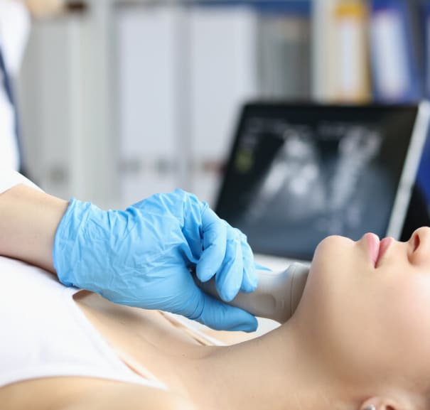
A lymph node ultrasound is a painless, non-invasive scan that uses sound waves to create images of the lymph nodes in your body. It allows your doctor to look at the size, shape, and structure of your lymph nodes without using any radiation.
Your doctor may recommend a lymph node ultrasound to help diagnose the cause of swelling, check for abnormal lymph nodes, or monitor an existing condition. It can also be used to help guide a biopsy needle if your doctor needs to take a sample of tissue for testing.
A lymph node ultrasound is especially useful in areas like the neck, where lymph nodes are close to the surface of the skin and easy to check.
Why Has My Doctor Recommended a Lymph Node Ultrasound?
There are many reasons why your doctor might suggest a lymph node ultrasound. You may have noticed a lump in your neck, armpit or groin, or perhaps your doctor picked something up during a routine exam. Other reasons include:
-
Persistent swelling in one or more lymph nodes
-
Pain or tenderness in the area of a lymph node
-
Unexplained weight loss, night sweats or fever
-
Signs of infection or inflammation
-
As part of a cancer diagnosis or treatment plan, such as for breast cancer
An ultrasound helps your doctor determine whether a lymph node looks normal, reactive (enlarged due to infection or inflammation), or potentially cancerous.
What Can a Lymph Node Ultrasound Diagnose?
Lymph node ultrasounds can help your doctor identify many different health conditions. These include:
-
Reactive lymph nodes: Enlarged due to a recent infection, but not cancerous. They usually appear oval with a healthy internal structure.
-
Lymphadenitis: Inflammation of the lymph nodes, often due to infection. These nodes may look swollen and have increased blood flow.
-
Lymphoma: A type of blood cancer that affects the lymphatic tissue. Nodes may appear rounder, darker (hypoechoic), and more vascular.
-
Metastatic cancer: Cancer that has spread to the lymph nodes from another part of the body. These nodes may appear uneven and have abnormal pockets of fluid or tissue.
-
Tuberculosis: May cause tissues within the lymph nodes to die (necrosis). They may look bubbly or congested.
-
Sarcoidosis: A condition that can cause clusters of enlarged lymph nodes.
-
Cat scratch disease: Usually results in large, tender lymph nodes near the site of infection.
-
Infectious mononucleosis: Often shows up as multiple enlarged, tender nodes, sometimes with areas of swelling.
-
HIV-related lymphadenopathy: May cause widespread lymph node enlargement.
-
Autoimmune diseases: Conditions like lupus or rheumatoid arthritis can lead to swollen lymph nodes with certain patterns.
While a lymph node ultrasound can’t always give you a definite diagnosis, it’s very useful for narrowing down the causes of your symptoms. It can also guide your doctor to offer further tests, such as a biopsy or a CT or MRI scan.
Types of Lymph Node Ultrasounds
There are several ultrasound techniques that your doctor can use, depending on your symptoms, where they are and what your doctor is looking for:
-
B-mode (grey scale): This is the standard type of ultrasound, showing detailed black-and-white images of the lymph nodes.
-
Doppler ultrasound: Measures blood flow within the lymph nodes. Cancerous nodes often have increased or abnormal blood flow.
-
Contrast-enhanced ultrasound: Uses special contrast agents or dyes to make blood vessels and structures within the lymph node clearer. This helps your doctor tell which nodes are reactive and/or cancerous.
-
Elastography: Measures the stiffness of a lymph node. Cancerous nodes are usually stiffer than benign ones.
-
Endobronchial ultrasound (EBUS): A specialised test to look at lymph nodes in the chest. Your doctor may use this type of ultrasound if they are worried your symptoms may be a sign of lung cancer.
-
Endoscopic ultrasound (EUS): Helps assess lymph nodes in the abdomen or near the digestive tract and helps your doctor to take tissue samples for testing.
How Does an Ultrasound of the Lymph Nodes Work?
An ultrasound scanner emits high-frequency sound waves from a small probe called a transducer. These sound waves bounce off tissues in the area of your body being examined, producing echoes that are processed by the ultrasound machine to create images.
What Equipment is Used During an Ultrasound?
Your sonographer will use some, or all, of the following equipment during an ultrasound scan:
-
Transducer: A small, handheld device that emits the ultrasound waves and then receives the returning echoes. It’s moved over the area of your body that your sonographer wants to check.
-
Ultrasound gel: This is applied to the skin to improve the transmission of sound waves between the transducer and your body.
-
Ultrasound machine: This processes the echoes received by the transducer and generates images on a monitor for your sonographer to look at.
-
Other tools: Depending on the procedure you’re having, for example, a biopsy, your imaging technician may use extra tools, such as needles or syringes.
What Are the Benefits of a Lymph Node Ultrasound?
If you’re unsure about having a lymph node ultrasound, it’s worth knowing all the benefits of the procedure, including:
-
It’s safe and gentle: A lymph node ultrasound doesn’t use any radiation and doesn’t hurt at all. It’s safe for almost everyone, including if you’re pregnant.
-
Gives a clear picture: It shows detailed images of your lymph nodes — including their size, shape and blood flow — so your doctor can spot anything unusual.
-
Can catch things early: If something’s not quite right, an ultrasound can help pick it up early. That means you can get the care you need as soon as possible.
-
Helps guide next steps: If your doctor needs to take a closer look with a biopsy, the ultrasound can help guide exactly where to take the sample.
-
Monitors your treatment: If you’re having cancer treatment, regular ultrasounds can help check how well it’s working and whether anything has changed.
-
It can help detect signs of infection or inflammation: such as infections like TB or conditions like glandular fever and arthritis that might be affecting your lymph nodes.
-
Supports diagnosis of lymphatic conditions: If you’re experiencing swelling or other symptoms, it can help doctors understand if a condition like lymphedema is involved.
-
Can check nearby blood flow: Sometimes it also gives useful information about nearby blood vessels, which can help assess your overall health and reduce risks.
What Are the Risks of a Lymph Node Ultrasound?
Lymph node ultrasounds are very safe because they don’t use radiation, but like any procedure, there are a few things to be aware of. You might feel a little pressure from the ultrasound probe, especially if the area is already tender, but it shouldn’t be painful.
If you’re having a needle biopsy guided by ultrasound, there’s a small risk of bruising, bleeding, or infection, but your medical team will take every precaution to keep you comfortable and safe.
Overall, an ultrasound is a low-risk scan that gives your doctor helpful information to guide your care.
Do I Need to Prepare for My Lymph Node Ultrasound?
In most cases, there’s nothing special you need to do beforehand. You can eat and drink as normal and take your usual medications.
You may be asked to wear loose-fitting clothing to make it easier to reach the area being scanned—especially if the scan is of your neck, armpits, or groin.
What Will Happen During My Lymph Node Ultrasound?
You’ll be asked to lie on an exam table or couch, often with your head turned or arm raised, depending on where you need the scan. Your sonographer will apply the ultrasound gel to your skin to help the transducer move smoothly and improve image quality.
The sonographer will move the transducer over the area to examine your lymph nodes. You might feel some gentle pressure, but it shouldn’t hurt.
In some cases, you may need Doppler imaging for a more detailed look at the blood vessels.
Your sonographer will look at the real-time results of the ultrasound and take images for review later on.
What Happens After a Lymph Node Ultrasound?
After the scan is over, you can wipe the gel off with tissue and get on with your day, unless you’re having further testing. Your images will be sent to your referring clinician for review.
When and How Will I Get My Results?
After your scan, the images are reviewed by a radiologist, a doctor who specialises in interpreting scans. They’ll write a report and send it to the doctor who referred you.
You’ll have a follow-up appointment with your clinician to go over the results. In some cases, your doctor might recommend further testing such as:
-
A lymph node biopsy
-
Blood tests
-
A CT or MRI scan
Remember, not all enlarged lymph nodes mean cancer. Most are benign and caused by infections or other harmless conditions. Your doctor will guide you through the next steps.
How Much Does a Lymph Node Ultrasound Cost?
Costs can vary depending on the clinic, where you live in the UK and the type of scan you’re having. In private clinics, you can expect to pay between £140 and £600 in the UK.
Private clinics often offer faster appointments and quick results. If you're having your scan through the NHS, it will be free, but you’ll usually have to wait longer for an appointment.
How Can I Get a Lymph Node Ultrasound?
You can ask your GP for a referral, or choose a private clinic and book directly via Scan.com. If you’re booking a private ultrasound, it’s important to choose the right scan for the area you’re worried about. Most lymph node ultrasounds are done on the neck, armpits, or groin. If you’re unsure, you can always check with our clinicians to make sure you’re booking the right one.
At Scan.com, we make it easy to find trusted providers near you – so you can book your private ultrasound scan quickly and confidently, with support at every step.
FAQs
What Does a Healthy Lymph Node Look Like on Ultrasound?
A normal lymph node is usually small, oval-shaped, and has a consistent internal pattern. It will often have a visible central area called the hilum, where blood vessels enter and leave the node.
What Does a Cancerous Lymph Node Look Like on the Scan?
If a lymph node looks unusual on an ultrasound, there are a few features that might raise concern. Things like a larger size, a rounder shape, or a darker appearance can sometimes point to cancer. The scan might also pick up changes like uneven texture, a missing fatty centre, thickened edges, or tiny calcium deposits.
In some cases, parts of the node may even show signs of damage or blurred borders if nearby tissue is involved. These signs don’t always mean cancer, but they help your doctor decide if more tests are needed to be sure.
Your radiologist may also check the blood flow in your lymph nodes using colour Doppler, which adds extra detail to your scan. Some patterns, like blood flowing mainly around the edges, or unusually high resistance, can sometimes suggest the node is under stress or packed with extra cells, which might happen with cancer. They’ll also look for unusual or misplaced vessels that don’t behave as expected. These findings aren’t always a cause for alarm, but they do help guide whether you need follow-up tests or scans.
What Will an Ultrasound of a Swollen Lymph Node Show?
A swollen lymph node on ultrasound may appear enlarged, and in many cases, it looks darker than the tissue around it (this is called hypoechoic). Depending on what's causing the swelling, the scan might also show increased blood flow, especially if your body is fighting an infection or if the node is cancerous.
How Long Does a Lymph Node Ultrasound Take?
The scan usually takes 15 to 30 minutes, depending on the number of lymph nodes to be checked and their location in your body.
Sources:
Cystic (necrotic) lymph nodes. (2021). https://radiopaedia.org/articles/cystic-necrotic-lymph-nodes?lang=gb
Lymph node biopsy guided by ultrasound. (N.D.) https://www.cancerresearchuk.org/about-cancer/tests-and-scans/lymph-node-biopsy-guided-ultrasound
Sonographic features of malignant lymph nodes. (2023). https://radiopaedia.org/articles/sonographic-features-of-malignant-lymph-nodes?lang=gb
The lymphatic system. (2024). https://lymphoma-action.org.uk/about-lymphoma-what-lymphoma/lymphatic-system
Ultrasound and X-ray scans. (2024). https://lymphoma-action.org.uk/about-lymphoma-tests-diagnosis-and-staging/ultrasound-and-x-ray-scans
Ultrasound-guided biopsy of the neck. (2023). https://www.qvh.nhs.uk/wp-content/uploads/2020/11/Ultrasound-guided-biopsy-of-the-neck-0175.pdf
Yu, S.C., et al. Necrosis in lymph nodes and their differential diagnoses: application of reticulin staining. (2024). https://pubmed.ncbi.nlm.nih.gov/37392241/
