
Ovary Ultrasound: Information & Get a Scan
An ovary ultrasound is a type of medical scan that uses high-frequency sound waves to produce detailed images and information on the size, shape and location of the ovaries. It will also show any abnormalities that need treatment or further investigation..
Carried out by a medical professional called a sonographer, doctors use the real-time images produced by an ultrasound to diagnose conditions affecting the ovaries.
In this article, we’re going to discuss in detail what an ovarian ultrasound is, what different types of ultrasound are available, why you might need one and what they can diagnose.
Key Takeaways:
-
The ovaries are two small, almond-shaped organs situated in the pelvic cavity that form part of the female reproductive system.
-
An ultrasound scan is carried out by a medical professional called a sonographer and uses high-frequency sound waves to create detailed images of the inside of the body. Quick, painless and minimally invasive, they’re used to give doctors detailed information on the health of the organs and soft tissues. The sound waves cannot be heard or felt as they enter the body.
-
An ovary ultrasound can be transabdominal (where an ultrasound probe is moved over the skin of the tummy) or transvaginal.
-
During a transvaginal ultrasound, your sonographer will pass a small, thin ultrasound probe into the vagina. It will be covered by a latex sheath and lubricating gel.
-
Doctors use the images created during an ovary ultrasound to diagnose and rule out a number of conditions that can affect the ovaries, including ovarian cysts, ovarian cancer and an ectopic pregnancy.
What is an Ovary Ultrasound?
The ovaries are a pair of organs, situated deep within the pelvic region, either side of the uterus (womb). Small and almond-shaped, they form part of the female reproductive system. They’re responsible for producing mature eggs and the female sex hormones oestrogen and progesterone and are connected to the uterus via two tubes called the fallopian tubes.
An ovary ultrasound scan is a routine medical procedure that uses sound waves to produce detailed, black and white images of each ovary. Due to the small size of the ovaries, their location and the fact that they can move within the pelvis, an ultrasound of the ovaries is usually carried out as both a transabdominal (tummy) ultrasound and a transvaginal ultrasound.
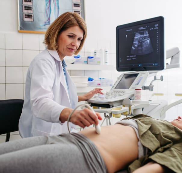
Transabdominal ultrasound scans are carried out externally. An ultrasound probe is moved over the skin of the abdomen and lower down over the pelvic area between the belly button and the pelvic bone.
During a transvaginal (or internal) ultrasound, your sonographer will place a small, thin ultrasound probe into the vagina to take images of the vaginal wall, cervix, uterus and ovaries from the inside of the vagina. The probe will be covered by a latex sheath or condom, as well as a lubricating and conductive gel, which will feel cold and wet.
Taking images both externally and internally ensures that the ovaries, as well as the fallopian tubes and the uterus are all scanned at the same time. This helps doctors diagnose and rule out conditions that can affect the ovaries, fallopian tubes and uterus that may have similar symptoms.
Both external ovary ultrasound scans and transvaginal ultrasounds are carried out on an outpatient basis. You’ll be able to return home the same day and once your results have been reviewed, you’ll be called by telephone back into the clinic to discuss your results and any recommended treatment and next steps.
Why You Might Need an Ovarian Ultrasound
Speak to your doctor or GP about being referred for an ultrasound scan of the pelvic region and ovaries if you’re experiencing any of the following symptoms, especially if accompanied by unexplained weight loss or extreme fatigue:
-
Pain in your lower abdomen or your lower left or right side
-
Lower back pain
-
Abdominal swelling or persistent bloating
-
Unusual vaginal bleeding
-
Pain during sex
-
Needing to urinate more frequently or more urgently
-
Changes in bowel habit
-
Feeling full very quickly and/or a loss of appetite
Your doctor may also recommend an ovary ultrasound scan if you’re having difficulty getting pregnant or your doctor suspects you may have an ectopic pregnancy.
What an Ovary Ultrasound Can Diagnose
Ovarian ultrasound scans can diagnose (and rule out) the following conditions:
-
Ovarian Cysts - fluid-filled sacs that form on or in the ovaries that are usually benign (non-cancerous)
-
Polycystic Ovary Syndrome (PCOS) - a condition that causes multiple cysts to develop within the ovaries
-
Ovarian Torsion - a condition that causes one or both of the ovaries to twist on its supporting fallopian tube and requires urgent medical attention
-
Endometriomas - dark-coloured cysts that form on the ovaries filled with tissue from the endometrium (lining of the womb), often associated with endometriosis (also known as ‘chocolate cysts’)
-
Ovarian Cancer - malignant (cancerous) tumours that have formed in the ovaries
-
Functional Cysts (Follicular or Corpus Luteum) - common cysts that can form on the ovaries during the menstrual cycle that often disappear without treatment
-
Hemorrhagic Cysts - common, blood-filled cysts that can form on the ovaries during ovulation and often disappear without treatment
-
Tubo-Ovarian Abscess - infected cysts affecting the ovaries and the fallopian tubes, often associated with untreated pelvic inflammatory disease that require urgent treatment
-
Dermoid Cyst (Mature Cystic Teratoma) - fairly common non-cancerous cysts that contain mature tissue from elsewhere in the body, including skin, hair and bone
-
Ectopic Pregnancy - a pregnancy that has started to develop in one of the fallopian tubes (instead of the lining of the womb) and requires urgent medical attention
Types of Ovarian Ultrasounds
There are a few different types of ovary ultrasound - which one you have will depend on your symptoms and medical history:
Transvaginal Ultrasound (TVUS)
A transvaginal ultrasound is performed using a thin ultrasound probe inserted into the vagina. It creates images of the ovaries and the rest of the female reproductive organs from inside the vagina. The probe is covered with a latex sheath or condom, and a gel is applied that lubricates the vagina and helps the sound waves travel into the body.
Transabdominal Ultrasound
A transabdominal ultrasound is performed using a standard ultrasound probe that’s moved over the skin of the abdomen and the pelvic region. A conductive gel is applied to the skin that will feel cold and wet.
Doppler Ultrasound
A Doppler ultrasound works in the same way as a transabdominal ultrasound, but it’s used to also create images of the surrounding blood vessels. Doppler ultrasounds create a “whoosh” sound as they detect blood moving through the blood vessels.
3D Ultrasound
3D ultrasound scans are carried out in the same way as a standard transabdominal ultrasound using slightly different technology to produce three-dimensional images of the ovaries. These more detailed images are used to monitor conditions such as small ovarian cysts to see if they’re growing and therefore require surgery to remove them.
Contrast-Enhanced Ultrasound
During a contrast-enhanced ultrasound of the ovaries, tiny microbubbles are injected into a blood vessel (they cannot be felt). As the sound waves meet the microbubbles, they create a more detailed image of the ovaries than a standard ultrasound.
How an Ultrasound of the Ovaries Works
An ultrasound scanner sends high-frequency sound waves into the body via special probes called transducers - they cannot be heard or felt as they enter the body. When sound waves enter the body, aided by a special conductor gel applied to the skin and/or the probe, they reach different tissues such as bones, organs and soft tissues.
As they encounter each different tissue type, the sound waves bounce back to the transducer probe, in the same way as an echo does. These echoes differ, according to the tissue type they’ve encountered. For example, they’re echoed back quickly from dense bone tissue, and more slowly from softer organs.
Ultrasound computers turn these echoes into images that are then reviewed by a radiologist.
Equipment Used
An ultrasound scanner is a small machine that sits on a medical trolley next to a bed. Transabdominal and transvaginal transducer probes are attached to the machine, along with a computer and a screen.
A clear gel is also used, either on the skin during a transabdominal scan or on the probe for a transvaginal scan. This gel acts as a lubricant to make the procedure more comfortable, and also as a conductor to help the sound waves penetrate the skin and echo back to the probe.
Benefits
Ultrasound scans are quick, effective and safe - they’re suitable for the majority of patients, including pregnant women. Also, unlike CT scans and x-rays that use ionising radiation, there’s no risk of radiation exposure from an ultrasound scan.
You can have a transvaginal ultrasound scan if you’re having your period, but let your sonographer know. If you’re wearing a tampon, it will need to be removed beforehand.
Risks & Side Effects
There’s no known risks or side effects to having an ovary ultrasound scan. However, if you’re experiencing abdominal or vaginal pain, it may feel uncomfortable, especially as you’ll need to have a full bladder for your appointment.
If you’re having a transvaginal ultrasound scan, the probe will be covered with a latex sheath. If you’re allergic to latex, let your sonographer know.
How to Prepare for an Ovary Ultrasound
There’s little preparation required for an ovarian ultrasound. Unless you’ve been told otherwise by your doctor, you can eat, drink and take medications as normal before your appointment.
You will however, need to have a full bladder as this helps to create the most beneficial images. Therefore, you’ll be asked to drink 500ml of water, half an hour before your appointment, without emptying your bladder.
The Procedure Explained: What to Expect
An ovary ultrasound is carried out in the following steps:
-
Check-In and Provide Medical History - When you arrive, your sonographer will ask questions about your symptoms and medical history and give you the opportunity to ask questions about your procedure.
-
Change Into a Gown (if needed) - For most ovary ultrasound scans, you’ll need to remove your lower clothing and underwear and change into a medical gown.
-
Lie Down on the Examination Table - Your sonographer will help you onto a bed. You’ll be lowered back to an almost lying position on your back, and if you’re having a transvaginal scan, your legs will be placed in stirrups. Your sonographer will cover your lower half in a blanket or tissue.
-
Apply Gel to the Abdomen (Transabdominal) - Conductor gel will be applied liberally to the skin of your tummy area so that the transducer can glide easily over the skin.
-
Insert Probe Into Vagina (Transvaginal) - A condom will be used to cover the transvaginal probe and the same gel will then be applied to the covered probe before it’s slowly placed inside the vagina.
-
Scan the Ovaries and Uterus - A transabdominal and a transvaginal scan are carried out separately, but at the same appointment. During each scan, your sonographer will move the probe, pause to take images and repeat until all images are taken.
-
Take Measurements and Images - They will also take measurements of the ovaries and the womb lining using the ultrasound computers.
-
Clean Up and Get Dressed - Once they have all the images they need, your sonographer will remove the probe and wipe away the gel. They’ll provide you with more tissue to wipe away any remaining gel and you’ll be able to get dressed.
What Happens After an Ovary Ultrasound?
Once your sonographer has taken the images they require, the scan will be over and you’ll be free to return home and to your normal duties.
Getting the Results
Your ultrasound images will be sent directly to a radiologist who specialises in reading ultrasound scans. Then, along with your referring doctor and the information provided by any other tests and scans you’ve had, they will review the images and make a diagnosis or rule out certain conditions.
Once reviewed, you will then receive a phone call to return to the clinic, and your results will be discussed with you. If necessary, your doctor will also discuss any treatments and further steps that may need to be taken.
How Much Does an Ovary Ultrasound Cost?
In the UK, you can expect to pay between £150 and £400 for a private ovarian ultrasound scan.
Get an Ovary Ultrasound
Having symptoms such as pain and abnormal vaginal bleeding or having fertility difficulties is worrying and stressful. Being referred for tests and scans to discover what might be causing your problems is important for getting treatment.
At Scan.com, we take the worry and stress out of waiting for an appointment. Book a private abdominal ultrasound with us today (this will include an ultrasound of your ovaries and uterus) and begin your journey to getting answers and starting the right treatment.
FAQs
Why is My Left or Right Ovary Not Visible on Ultrasound?
Excess gas or stool from the bowel can obscure either ovary, or it may be hidden behind another organ such as the bowel or bladder. In post-menopausal women, the ovaries can shrink as they no longer contain follicles or eggs, and can therefore be more difficult to view on an ultrasound.
What Does an Ovary Look Like on an Ultrasound?
On an ultrasound, the ovaries appear as small, almond-shaped structures. If you’re pre-menopausal, they will contain follicles - small sacs that each contain one egg that’s released during ovulation. These follicles appear as small, round, white structures on an ultrasound scan.
How to Tell Which Ovary Conceived on Ultrasound?
In early pregnancy (typically before 10 weeks) an ultrasound can pick up an empty follicle called a corpus luteum in either the left or right ovary. If an ovary contains a corpus luteum, it will suggest that it was this one that produced the egg that subsequently became fertilised.
How to Read Ovary Ultrasound?
Your radiographer and specialist doctors are trained to read and interpret your ovary ultrasound results and to diagnose or rule out medical conditions that might be affecting your ovaries.
How Long Does an Ovary Ultrasound Take?
Ultrasound scans of the pelvic area, including the ovaries and uterus, take between 15 and 30 minutes. Your appointment will take around an hour, including the time taken to get undressed and for any paperwork to be completed.
References
Ultrasound Scans for diagnosing ovarian cancer. (2024, June 25). Ovarian Cancer Action. https://ovarian.org.uk/ovarian-cancer/ovarian-cancer-diagnosis/ultrasound-scans/
Pelvic ultrasound scan. (n.d.). Cancer Research UK. https://www.cancerresearchuk.org/about-cancer/tests-and-scans/ultrasound-pelvis
Professional, C. C. M. (2025, March 19). Pelvic Ultrasound. Cleveland Clinic. https://my.clevelandclinic.org/health/diagnostics/4997-pelvic-ultrasound
Website, N. (2023, June 28). Ovarian cyst. nhs.uk. https://www.nhs.uk/conditions/ovarian-cyst/
