
Ankle X-Ray: Info and Get a Scan
If you’re experiencing pain, numbness, tingling or stiffness in your ankle joint, or it feels tender to the touch, you may be concerned that you have a broken or fractured ankle. Or you may be worried that another underlying condition, such as arthritis, is causing your joint stiffness.
An ankle X-ray can be hugely helpful in diagnosing fractures and breaks in ankles, as well as conditions such as arthritis and gout. It can help you get the answers you need so you’ll be back on your feet sooner. We’ll guide you through what an ankle X-ray involves, when you might need one and how it can help your doctor diagnose trauma or injury to the area or underlying conditions that may be causing pain, stiffness or weakness in the joint.
Understanding ankle X-rays
An ankle joint X-ray is an imaging tool doctors use to see what’s happening inside your ankle. It uses small doses of radiation to take images of the tiny bones and soft tissue within the joint so that your doctor can identify any injuries or fractures. They can also check for other problems, such as joint deformities in the ankle, and signs of underlying conditions, such as arthritis or gout, that can affect the ankle joint, causing pain, inflammation, weakness or stiffness.
Ankle X-rays are quick and usually painless. However, your technician may have to move and position your foot to allow X-ray of ankle images to be taken from different angles, which may feel uncomfortable. But they will take great care not to hurt you, especially if they suspect you have a broken or fractured ankle.
The ankle X-ray image will be black and white with shades of grey in between. Bones will appear white because their density blocks more X-rays from passing through them, while skin, muscles and ligaments will appear in shades of light grey to black.
Ankle X-ray anatomy: What you need to know
Knowing which bones are imaged during an ankle X-ray can be an enormous help in understanding what the X-ray can reveal. Here’s a quick glossary of key terms:
-
Tibia: The larger, weight-bearing bone of the lower leg, also known as the shinbone.
-
Fibula: The smaller bone that runs parallel to the tibia, providing lateral stability.
-
Talus: The bone that connects the lower leg to the foot, forming the ankle joint.
-
Joint spaces: The gaps between the bones. These should appear uniform; any narrowing or widening can be a sign of issues like arthritis or ligament injuries.
When to get an ankle X-ray
If you’ve injured your ankle or are worried about an underlying condition affecting the joint, it’s important to know when you might need an X-ray. Here are the key signs that could signal when to X-ray an ankle injury:
-
You can’t put weight on your injured ankle or take a few steps without pain.
-
You’re experiencing pain or tenderness on the outer or inner ankle bone.
-
You feel tenderness at the base of the fifth metatarsal (the bone on the outer side of your foot) or the navicular bone (in the midfoot).
-
Your pain is intense, or if there’s noticeable swelling or bruising around the ankle.
-
Your ankle looks deformed or misaligned.
-
Your symptoms have not improved after a few days of rest, ice, compression, and elevation (RICE).
-
You have ongoing joint pain that doesn’t improve with rest or over-the-counter pain relievers.
-
You notice swelling, tenderness, or stiffness in your joints, especially in the morning or after periods of inactivity.
-
You see visible changes in the shape of your joints over time.
-
You may be experiencing a decreased range of motion or finding it harder to perform your usual daily activities because of discomfort.
-
You have a family history of arthritis or related conditions and are concerned about your ankle joint.
-
You have sudden, severe pain in a joint (often the big toe) that feels like a burning sensation.
How do I know if I need an X-ray on my ankle or foot?
Your clinician may recommend that you have a more comprehensive foot X-ray rather than an ankle X-ray, based on their physical examination of your ankle and foot. Based on your symptoms, they use a particular guideline to work out when an ankle or foot X-ray is more appropriate: the Ottawa Ankle Rules. Here’s a simple breakdown of how they do it:
Ottawa Ankle Rules
Your doctor will recommend an ankle X-ray if:
-
You have bone tenderness at the posterior edge or tip of the lateral malleolus (outer ankle bone).
-
You have bone tenderness at the posterior edge or tip of the medial malleolus (inner ankle bone).
-
You cannot bear weight for four steps, on the affected side, immediately after the injury and during the physical examination.
Your doctor will recommend a foot X-ray if:
-
You have bone tenderness at the base of the fifth metatarsal (the bone on the foot's outer side).
-
You have bone tenderness at the navicular bone (this is in the midfoot).
-
You are unable to bear weight for four steps (as above).
Following these rules means that your clinician is unlikely to recommend a more expensive foot X-ray and unnecessary imaging unless they think it is necessary.
When should you get an X-ray for a sprained ankle?
If you have mild swelling and pain without tenderness and can bear weight on the injured ankle, an X-ray may not be necessary. However, it depends on the severity of your symptoms. That’s why doctors use the Ottawa Ankle Rules to be sure they’re recommending the proper imaging technique for your symptoms.
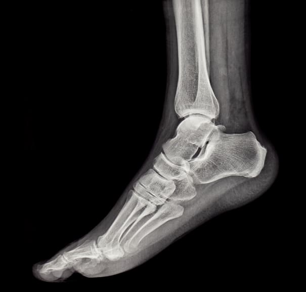
Normal ankle X-ray vs. fracture
A normal ankle X-ray shows a clear view of the ankle bones, including the tibia, fibula, and talus bones. The tibia is the shinbone which runs from the knee to the ankle. The fibula is the smaller bone of the lower leg, which runs parallel to the shinbone. The talus is a small bone which sits just above the heel bone. There should be no signs of fractures or abnormalities to the bones in a normal vs fracture X-ray. The spaces within the joint should appear normal, and there should be no swelling or bone displacement.
What does a broken ankle look like on X-ray images?
In contrast, a fractured ankle X-ray will reveal clear signs of injury. You may see breaks or cracks in the bones, misalignment, or fragments where the bone has been displaced. A broken ankle X-ray will also show swelling around the joint, indicating soft tissue injury.
What will an X-ray of the ankle show?
While X-rays are valuable tools in helping doctors identify injuries and many underlying conditions, here’s what X-rays of the ankle can and can’t reveal:
Healthy ankle X-ray
A healthy ankle X-ray will show bones with no fractures or deformities. The joint spaces are well-defined, with no signs of swelling or abnormalities.
Avulsion fracture ankle X-ray
An avulsion fracture happens when a ligament or tendon pulls off a small piece of bone. On an X-ray, this appears as a tiny bone fragment near the main bone.
Sprained ankle X-ray vs. normal ankle X-ray
An X-ray won’t typically show a sprain since it primarily affects the ligaments, not the bones, and X-rays are not the most suitable imaging technique for showing soft tissue damage. However, your doctor may recommend an X-ray to rule out a fracture and to be on the safe side, or an MRI to check the ligaments.
Torn ligament in ankle X-ray
If you’re wondering can an X-ray show torn ligaments in the ankle, the answer is no - they only reveal bone injuries. If your doctor suspects a torn ligament, they may recommend alternative imaging like an MRI or an ultrasound.
Severe arthritis in ankle X-ray
An X-ray of severe arthritis will show narrowing of the joint space, bone spurs, and possible bone deformities. It helps your doctor assess how much the condition has damaged your joints.
Gout ankle X-ray
In chronic gout, X-rays may show urate crystal deposits caused by the condition, bone erosion, and narrowing of the joint spaces. If you are in the early stages of having gout, it might not yet be visible on an X-ray.
Dislocated ankle X-ray
A dislocated ankle X-ray will reveal any bones out of their normal alignment, often with the joint visibly displaced.
Ankle X-ray views
During your ankle X-ray, your technician will take lots of pictures of your ankle from different angles. These images will help to provide a complete view of the bones and joint structure, which is crucial for your doctor to diagnose a fracture or other injury.
Here are the views your technician will take during the ankle anatomy X-ray.
-
Anteroposterior (AP) view: This is the front view of the ankle, showing the alignment of the tibia, fibula, and talus bones. It helps your doctor check the overall structure and alignment of your ankle.
-
Lateral view: The side view of the ankle, known as the lateral ankle X-ray, gives your clinician a detailed view of the ankle joint, including the tibia, fibula, talus, and calcaneus (heel bone). This view is essential for identifying fractures and dislocations and checking the joint space between the bones.
-
Internal oblique or mortise view: This angled view is taken with the foot rotated slightly inward to get a better picture of the ankle joint. It helps your doctor get a clearer picture of the space between the tibia and talus and the distal fibula, helping to identify fractures or joint space narrowing in the case of arthritis. It’s particularly useful for detecting injuries that might not be visible in the AP or lateral views.
Ankle X-rays are key in diagnosing issues like fractures, dislocations, arthritis, and gout. They provide clear images from multiple angles (front, side, and angled) to help your doctor see exactly what’s going on inside your ankle. While X-rays are great for showing bones, they don’t reveal ligament damage, so your doctor might suggest additional tests if needed.
If you’re unsure whether you need an ankle X-ray or a different imaging technique, such as an MRI or an ultrasound, our friendly clinicians can help guide you in the right direction during a consultation, and there’s no obligation to have an X-ray or scan afterwards. Fast-track your diagnosis with no waiting lists and get the treatment you need sooner. Book your ankle X-ray today with Scan.com.
Sources:
Ankle radiograph (an approach). (2024). https://radiopaedia.org/articles/ankle-radiograph-an-approach?lang=gb
How should I assess a suspected sprain or strain? (2020). https://cks.nice.org.uk/topics/sprains-strains/diagnosis/assessment/
Ottawa ankle rules. (2018). https://radiopaedia.org/articles/ottawa-ankle-rules-1?lang=gb
X-Ray Exam: Ankle. (2022). https://kidshealth.org/en/parents/xray-ankle.html
