
Hand X-ray: Information & Get a Scan
X-rays are quick and painless, and are used to diagnose or rule out many different medical conditions. They can be used to scan most body parts, including the hands.
What is an X-ray?
An X-ray is a type of medical scan that takes black and white images of the inside of the body, without having to operate or perform invasive procedures. X-rays show the bones, which appear white on an X-ray image, and soft tissues, organs, fat and muscles, which appear darker. They clearly show the structure of the joints and soft tissues, and highlight any abnormalities such as breaks, tears and inflammation.
An X-ray uses a small amount of radiation that passes through the body. This radiation cannot be seen or felt.
Different tissues absorb x-rays at different rates - for example, x-rays cannot pass through bones as well as they can through soft tissues, therefore, the bones show as white on an x-ray image. X-rays can pass easily through organs, fat, muscle, tendons and ligaments and these show darker, in shades of black and grey on an X-ray.
What is a Hand X-Ray?
A hand x-ray focuses on one or both of your hands and may include your wrists. It helps doctors diagnose a range of conditions that may be causing pain, stiffness or problems with movement, as well as conditions that might be causing visible lumps or changes in the shape of your hands, fingers or thumbs.
Who Performs a Hand X-ray?
A hand x-ray is usually performed by a trained medical professional called a radiographer in the radiography department of your hospital or clinic.
What a Hand X-Ray Can Diagnose
Hand x-rays are routinely used to diagnose a wide range of conditions, including bone breaks, soft tissue injuries and diseases such as arthritis and gout.
Fractures
Bone breaks, or fractures, in the hand can be caused by sports or workplace injuries and trips and falls where you put your hands out to break your fall. They can affect one or more of the many bones in your hand and will cause immediate pain and swelling.
Dislocations
A dislocation, where a bone is forced out of its normal position, can again be caused by accidents, trips and falls, and can affect any of the bones within the hand and wrist joint. They too cause immediate pain, and you may see that the affected bone, usually in the finger or wrist, looks visibly out of place.
Arthritis
Osteoarthritis (caused by wear and tear) and rheumatoid arthritis (an autoimmune disease where the immune system mistakenly attacks the joints) are both types of arthritis that can affect the hand joints. Both cause a wearing away of the cartilage that cushions the joints (leading to pain, stiffness and immobility) that can be seen on x-ray images.
Osteoporosis
Osteoporosis, or “brittle bones”, can lead to a weakness in the bones that will mean a higher risk of bone fractures. A hand x-ray will show bone breaks and fractures, but a DEXA x-ray is used to specifically diagnose osteoporosis.
Bone Infections
A bone infection, called osteomyelitis, can develop in the bones of the hand and results in pain, swelling, redness, heat and inflammation that gets worse over time. Osteomyelitis in the hand can be caused by an infected wound or develop after hand surgery or it can spread to the hand in the bloodstream from elsewhere in the body.
Bone Tumours
Tumours in the bones of the hand are rare and are most commonly non-cancerous, or benign. An X-ray would normally be used alongside another scan such as an MRI or CT scan to diagnose a bone tumour and to determine if it’s benign or malignant (cancerous).
Growth Plate Injuries
The growth plate is an area of cartilage near the end of a bone, where new bone cells are formed allowing for the growth of the bone. Young children can injure the growth plates in the fingers and hand, that may cause the hands to develop growth or shape abnormalities.
Carpal Tunnel Syndrome
Carpal tunnel syndrome is caused by a pinching of a nerve that runs through the wrist into the hand, fingers and thumb, leading to tingling, pain and numbness in the palm and digits. It can be caused by injury and repetitive motions such as those through work or sports. An X-ray cannot diagnose carpal tunnel syndrome alone, but it can be helpful in ruling out other possible causes of symptoms.
Gout
Gout is a form of arthritis and causes extreme and often sudden pain in the joints, usually in the feet and toes but sometimes in the hands. It’s usually accompanied by redness, heat and swelling around the painful area. It’s caused by the buildup of a substance called uric acid, which forms painful crystals in the joints.
Congenital Bone Abnormalities
Congenital bone abnormalities are those that someone might be born with. In the hand, this can result in extra digits or fused fingers and thumbs. An X-ray can be helpful in guiding what corrective surgery may be possible.
Types of Hand X-Rays
There are various ways that a radiographer can perform a hand X-ray - which one you have will depend on your symptoms and medical history.
AP (Anteroposterior) View
Your hand is placed with your palm facing upwards to show the alignment of the hand bones, particularly those of the fingers, from the palm of your hand.
PA (Posteroanterior) View
Your hand is placed with your palm facing down to show the bones and their alignment from the top of your hand.
Lateral View
Your hand is x-rayed from the side, usually to assess the bones from a different angle in conjunction with x-rays taken from the AP and PA views. Taken together, these three views are helpful in diagnosing fractures and dislocations.
Oblique View
Your hand is positioned palm down, slightly rotated upwards so that the thumb is raised and the little finger rests on the digital recording plate. This view is helpful in diagnosing arthritis and complex fractures.
Ball-Catcher’s View (Nørgaard View)
Your hand is x-rayed whilst in the position of holding a tennis ball, which is helpful in the diagnosis of arthritis.
Stress View
Both hands will be placed in various stress positions, such as forming a diamond shape or pressing both thumbs together to help diagnose ligament or joint damage.
Flexion and Extension Views
Your hand or hands will be x-rayed with your hand relaxed then extended, and the results compared, which is helpful in diagnosing arthritis.
How a Hand X-Ray Works
X-rays work by passing a small amount of radiation through the body. This radiation creates energy, and the energy is absorbed by the bones and soft tissues of the hand.
During a hand x-ray, your hand will be rested on a digital recording plate that picks up the differences in energy absorption between the bones, tendons, ligaments, muscles and fat, and turns it into an image
Benefits
An X-ray is a quick, painless way of diagnosing conditions that might be causing pain, stiffness and immobility in your hands, wrists or fingers. They’re suitable for most people and only carry small risks.
Risks & Side Effects
There are no known side effects to having an X-ray, but there is a small risk of radiation exposure. Radiation naturally exists in low levels in the environment, and it’s called background radiation. Having an X-ray is thought to expose a patient to around the same amount of background radiation as being in the environment for between a few days to a few years, depending on the environment.
Having multiple X-rays throughout life will expose you to more radiation, but if the benefits of a diagnosis outweigh the risks, it’s considered the best course of action to have an X-ray. However, unless it’s absolutely necessary, it’s advised that pregnant women and young children avoid having an X-ray.
How to Prepare for a Hand X-Ray
A hand x-ray requires very little preparation. Unless advised otherwise, you can continue to eat and drink as normal beforehand, and carry on taking any regular medications. You will be asked to remove any metal jewellery and watches before entering the x-ray room, and it’s helpful if you wear a loose fitting or short sleeved top.
The Procedure Explained: What to Expect
Having a hand X-ray is a relatively quick process, taking around half an hour.
Change Into A Gown
When you arrive, you will be asked to leave all metal objects, including coins, bank cards, mobile phones and potentially any clothes containing metal such as a belt or a bra in a safe place. If you’re wearing a top that does not expose your wrist and hand, you’ll be asked to change into a gown.
Sit Or Stand In The X-Ray Room
Next, your radiographer will ask you to sit or stand near the X-ray machine, so that they can help you into the right position. They will be mindful that your arm or hand may hurt, and will take this into account when positioning you.
Position Hand On The X-Ray Plate
They will then ensure your hand is in the correct position on the digital recording plate for the type of hand X-ray you’re having.
The X-ray is Taken
Once you’re in position, you will be asked to remain very still. Your radiographer will move behind a lead screen where they can control the X-ray machine remotely. They will take an X-ray, and if necessary, re-position you for another.
Image Review
Once they’re happy that they have the correct and clear images, the X-ray will be over. The images will be sent to a specialist doctor called a radiologist to review. They are trained in spotting abnormalities and in diagnosing certain conditions.
They will then speak to your referring doctor and, if necessary, another doctor such as a bone specialist or orthopaedic surgeon, in order to confirm their diagnosis and formulate a treatment plan. Your medical team will then get in touch with you to discuss your results.
X-Ray Equipment
An X-ray machine is a large piece of medical equipment, housed in its own room. Depending on the type of x-ray you’re having, you may lie down on a bed, stand against a wall, or in the case of a hand x-ray, your hand and arm will be placed against the digital recording plate.
Your radiographer will be in the same room as you, and they will help you into the right position. But to take the X-ray, they will stand behind a lead-lined partition to reduce their radiation exposure. If you need multiple different hand x-rays, they will come out from behind the partition to help you into position, before returning again to take the x-ray.
What Happens After a Hand X-Ray?
After your hand-ray, you’ll be able to get dressed if you need to remove any clothing and leave the x-ray room. Your radiographer will give you any information you need, and you can return to your normal duties if you’re able to.
Getting the Results
It may take several days for your medical team to analyse your results and decide on the next course of action. This may be further tests and scans, or a diagnosis and treatment plan. In the case of a broken or dislocated bone, you’ll get your results the same day, and your medical team will decide how to proceed with surgery or a cast or both. Either way, your medical team will support and advise you throughout.
Costs
A private hand x-ray in the UK will cost between £55 and £150, depending on the type of x-ray you need. Consultations, follow ups and treatments will cost extra.
Get a Hand X-Ray
If you think you may need a hand x-ray, you may have to wait several weeks or months for one on the NHS if it isn’t an emergency. A private X-ray is usually much quicker. Book yours today and we’ll support you every step of the way.
FAQs
What Does Arthritis Look Like On An X-Ray Hand?
A hand x-ray will show arthritis as a narrowing of the gap between the affected bones, due to the disappearance of the cushioning cartilage layer.
What Does A Normal Hand X-Ray Look Like?
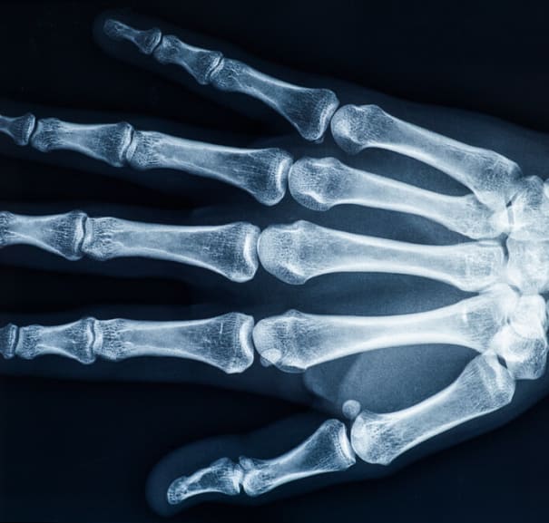
A normal hand x-ray will show, in white, the bones of the digits, palm and wrist in alignment and the healthy soft tissues, ligaments and tendons as grey and black, with no signs of inflammation or swelling.
How Long Does A Hand X-Ray Take?
A hand x-ray will take around 30 minutes - sometimes more if you require multiple different x-rays or your hand is injured and is painful or difficult to move.
What Does A Hand X-Ray Show?
A hand x-ray shows all of the individual bones of the fingers, thumb, hand and wrist, plus the surrounding soft tissues. It will highlight any abnormalities present in the bones and soft tissues.
References:
Professional, C. C. M. (2025b, February 19). Hand X-Ray. Cleveland Clinic. https://my.clevelandclinic.org/health/diagnostics/23518-hand-x-ray
Website, N. (2022c, May 4). X-Ray. nhs.uk. https://www.nhs.uk/conditions/x-ray/
Murphy, A., & Shetty, A. (2014). Hand series. Radiopaedia.org. https://doi.org/10.53347/rid-29804
Roth, E. (2017, March 15). Hand X-Rays. Healthline. https://www.healthline.com/health/hand-x-ray
