Key takeaways:
-
Knee pain can start suddenly following an accident or injury, or it can begin for no obvious reason and gradually get worse
-
Knee problems can affect the bones of the knee joint, the cartilage or the soft tissues (including the muscles, tendons and ligaments)
-
X-rays, MRI, CT and ultrasound scans are used to diagnose the cause of knee pain
-
The best scan for knee pain will depend on your symptoms and when they started
The causes of knee pain include infection, inflammation, general wear and tear and specific knee injuries. A knee scan provides detailed images of the knee joint and the structures around your knee that doctors can use to diagnose and plan treatment.
When Do You Need A Scan For Knee Pain?
Knee pain and discomfort can prevent you from moving around properly and doing the activities, sports and exercise you enjoy. A diagnostic scan provides clarity on what’s causing your pain and what treatment you need. Knee problems can be caused by accident, injury, overuse and wear and tear, and include:
-
Pain ranging from a dull ache in the knee area in general, to a sharp, intense pain in a specific part of the knee
-
Pain when moving the knee or at rest
-
Swelling, heat, redness and inflammation
-
Stiffness or weakness in the knee joint
-
Immobility or a feeling that the knee joint is “locked”
-
Feeling like your knee may give way when you put weight on it
-
Popping, clicking or grinding noises when moving the knee
If you’re experiencing any of these symptoms and your knee pain isn’t improving with rest, ice and over-the-counter painkillers, getting a scan to understand what’s causing your symptoms is the first step toward the treatment you need.
How is Knee Pain Diagnosed?
If you have knee pain, your doctor may perform an initial consultation and then organise one or more tests and scans to help diagnose the cause of your symptoms.
-
Physical Examination: Your doctor will ask you some questions about your knee problems, how long you’ve had them and whether anything makes them feel better or worse. They’ll then ask you to lie on an examination table while they feel around your knee. They may ask you to stretch and bend your leg to assess your range of motion and look for anything unusual.
-
Blood Tests: You may need some blood tests to check your inflammation levels. Erythrocyte sedimentation rate (ESR) and C-reactive protein (CRP) blood tests are routinely used to detect high levels of inflammation that could be causing your knee pain. A rheumatoid factor (RF) blood test helps diagnose rheumatoid arthritis, and a uric acid test helps detect gout. A complete blood count (CBC) will detect signs of infection. Blood tests alone cannot diagnose the exact cause of your knee problems, as they may detect infections or inflammation elsewhere in your body, but they’re useful when used in conjunction with other tests.
-
Joint Aspiration: If your knee is swollen, it may mean extra fluid has accumulated in the joint, causing pain. A joint aspiration is a quick, minimally invasive procedure that helps to relieve swelling and is also used to diagnose certain knee conditions. During a joint aspiration, your doctor will apply a local anaesthetic and then use a long, thin needle to draw excess fluid from the knee joint. This fluid will then be sent to a laboratory to check for signs of infection, bleeding and crystals.
-
Medical Imaging Scans: Diagnostic tests such as blood tests and joint aspiration will help identify potential causes of your knee pain, such as infection or inflammation. However, a scan of the knee joint will give your doctor a detailed view of the bones, muscles, cartilage, tendons and ligaments of the entire knee joint. From this, they can diagnose fractures, tendon and ligament injuries, cysts, soft tissue swelling and cartilage damage. Various knee scans are used to guide and plan treatment.
-
Diagnostic Arthroscopy: A knee arthroscopy is a type of keyhole surgery performed under a general anaesthetic to remove damaged tissues and loose pieces of cartilage that have been identified on a scan and may be causing pain. An arthroscopic procedure can also be used to obtain tissue samples (biopsies) from inside the knee joint if other tests and scans have proved inconclusive. These biopsies are tested in a laboratory for signs of infection or cancerous cells.
Once the cause of your knee pain and discomfort has been diagnosed, you can make informed decisions on your treatment, which may involve rest, anti-inflammatory medications, physiotherapy or a surgical procedure.
Types of Scans for Knee Pain
Having a scan to diagnose the cause of knee pain is a quick and reliable way to get answers. There are several types of knee scans:
X-Ray
An X-ray of the knee is often the first scan used to detect the cause of knee pain. X-rays use low-dose radiation to produce black and white static images of the knee joint, and they’re particularly useful for highlighting bone fractures, dislocations and abnormalities in the alignment of the knee joint.
MRI Scan
MRI stands for magnetic resonance imaging, and a knee MRI scan uses radio waves and a strong magnetic field to create detailed images of the inside of the knee joint and the tissues and structures around your knee.
Often considered the gold standard imaging test for knee injuries, an MRI scan is used to diagnose conditions affecting the bones, joints, soft tissues (including muscles, tendons and ligaments), nerves and blood vessels. They’re particularly useful for evaluating soft tissue injuries and joint strains and sprains.
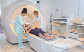
An MRI scanner is a large, doughnut-shaped machine. During an MRI scan, you’ll be required to lie on a bed that slowly moves into the MRI machine, while you lie completely still.
Patients with claustrophobia or who struggle to remain still may find having an MRI scan difficult. However, some clinics use an open-style MRI scanner that feels less closed in. Either way, for a knee scan, you’ll enter the scanner feet first, and your upper body and head may not need to enter the machine.
Unlike X-rays and CT scans, MRI scanners don’t use radiation. However, due to the strong magnets used, they’re unsuitable for patients with metal implants, meshes, clips, stents or prostheses.
MRI scans take longer than CT scans, but they may detect subtle bone fractures and abnormalities that CT scans might miss.
CT Scan
CT stands for computed tomography, and a CT scan uses radiation in the form of X-rays to create three-dimensional, cross-sectional images of the inside of the body.
Doctors use CT scans to diagnose conditions affecting the bones, soft tissues and blood vessels within the knee joint, often in emergency situations, such as following a car or motorbike accident. They’re particularly useful for assessing bone fractures that might not be detected on a standard X-ray.
A CT scanner is generally smaller and less claustrophobic than an MRI scanner, and because they don’t use magnets, they’re safe if you have metal implants or metallic shards or shrapnel in your body.
CT scans are quicker than an MRI scan, and involve lying on a medical table while the CT scanner moves around your knee to create a three-dimensional image of the joint. They use X-ray radiation in low doses.
Ultrasound Scan
Ultrasound scans use high-frequency sound waves to create real-time, moving images of the knee joint. Because they create dynamic, moving images, they’re useful for highlighting problems in which soft tissues catch on nearby bone, causing pain and clicking noises in the joint.
Diagnostic ultrasound scans are also used to detect cysts and soft-tissue inflammation.
Bone Scan
A bone scan is a type of nuclear medicine scan. During a nuclear medicine scan, a safe radioactive chemical called a tracer is injected into a vein, usually in the arm. The tracer emits radioactive gamma rays, which can be detected by a special camera used to image the knee.
Nuclear medicine bone scans are used to detect tumours in the bones and diagnose bone cancer. They’re normally only used if your doctor suspects you may have bone cancer that’s causing knee pain, or if everything else that could be causing your knee problems has been ruled out.
PET Scan
PET stands for positron emission tomography and is another type of scan that uses a radioactive tracer to highlight possible tumours in the bones and soft tissues.
Which Scan is the Most Effective?
Your doctor will recommend the best scan for your knee pain based on your symptoms and how they present:
-
For a First-Line Scan: an X-ray is often the first scan performed on a painful knee to identify or rule out areas of inflammation, bone fractures, dislocated kneecaps or gaps within the joint that could indicate worn cartilage.
-
For Ligament Injuries: MRI scans are used to detect ligament damage, tears and sprains.
-
For Meniscus Tears: an MRI scan can detect tears in the meniscus, the cartilage that cushions the knee joint and adds stability.
-
For Tendon Problems: MRI scans are used to detect problems affecting the knee tendons, including strains and tendonitis.
-
For Knee Swelling: an ultrasound scan is often used to confirm fluid buildup that’s causing swelling and bursitis (inflammation and swelling in the bursa, fluid-filled sacs that cushion the joints).
-
For Cartilage Damage: MRI scans are best for detecting cartilage damage.
-
For Early Arthritis Detection: an X-ray can detect general wear-and-tear in the knee joint, specifically areas where cartilage has been worn away, which is indicative of early osteoarthritis.
-
For Stress Fractures: an MRI is best for detecting small stress fractures in the bones of the knee joint.
-
For Persistent Unexplained Knee Pain: an MRI scan can be used to diagnose knee pain when there’s been no obvious injury or cause, by highlighting problems in the bones, soft tissues and cartilage.
-
For Suspected Infection: an X-ray is often the first scan used to diagnose unexplained knee pain and can highlight suspected areas of infection, especially in the bones. Alternatively, an MRI scan can detect infections in bones and soft tissues caused by septic arthritis.
-
For Tumours or Bone Lesions: specialist nuclear medicine scans, such as bone and PET scans, are used to detect tumours and cancerous lesions in bones and surrounding structures.
Get a Scan
Knee pain can affect your day-to-day life. Get answers fast with a private knee scan. Each booking is reviewed by our team, and if they think you may benefit more from a different scan to the one you booked, they’ll call you to discuss your options.
Affordable and fast, you’ll have your results in days. You can then choose to continue your care with one of our experts, or take your scan report and images to your NHS GP.
References
Website, N. (2025e, December 4). Knee pain. nhs.uk. https://www.nhs.uk/symptoms/knee-pain/
Professional, C. C. M. (2026, January 26). Knee pain. Cleveland Clinic. https://my.clevelandclinic.org/health/symptoms/21207-knee-pain
Al-Ani, Z. (2023, April 11). Knee pain; choosing the right scan! MSK US Injections. https://www.mskultrasoundinjections.co.uk/post/knee-pain-choosing-the-right-scan